By the way, if you are interested in reading about the less complex tendinous landmarks of the ventral forearm, check out the very first post, The Ventral Forearm: What are those Tendons?
So. When we last left off in our forearm saga, we were looking at, among other structures, the "twin muscles," two muscles that look very much alike and run directly down the dorsal side of the forearm. Their similarity to one another as well as their central location on the dorsal forearm make them among the easiest to identify in this area.
The last three muscles we'll cover on the dorsal forearm can be found just radial to the twins-- meaning closer to the thumb side of the arm compared to the centrally located twins. It's no coincidence that each of these three muscles have the root "radial" in their names. As well as indicating that these muscles are found on the radial side of the arm, this root also tells us that these muscles pull the hand toward that side. This movement is known as abduction of the hand. Hence the fact that radial side arm muscles tend to abduct.
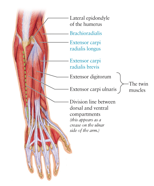
The three muscles we'll be looking for today are labeled in blue on this diagram. Notice that they are closest to the radial (thumb) side of the hand. And notice that all their names have "radial" somewhere in the name.
Of these muscles, the most radial is brachioradialis. This muscle starts way up on the upper arm (or brachium, hence the root brachio in its name) and travels distally, along the radius, towards its insertion on the styloid process of the radius (a bump at its distal end.) This muscle, despite its certified membership in the dorsal forearm compartment, can actually be seen more clearly from the ventral side. So we won't see much of it today in our photographs.
Running right between brachioradialis and extensor digitorum (one of the twin muscles) we see two muscles with very similar names: extensor carpi radialis longus and extensor carpi radialis brevis. Can you guess, by looking at these names, what these muscles have in common and what they don't?
Let's look at their names: Both names contain extensor carpi radialis, which means extensor of the wrist (carpi) on the radial side of the arm (radialis.) So these are attributes of both muscles. But how do we distinguish them from one another? The qualifiers tacked on to the end of each name tell us! Yes! One of these muscles is longer than the other. Extensor carpi radialis longus is the longer of the two, and extensor carpi radialis brevis is the shorter. (Brevis is Latin for short, or brief.) Knowing this, you should be able to tell one of these muscles from the other, as one is clearly longer than the other. In addition, it's helpful to know that extensor carpi radialis brevis lies right next to extensor digitorum and it tends to sink in rather than stand out when the dorsal forearm muscles show on the surface of the body.
Extensor carpi radialis longus is sometimes identified by its unique shape. It originates just proximal to the lateral epicondyle of the humerus, higher up on the arm than the origin points of the twin muscles. Also, unlike the twin muscles, extensor carpi radialis longus take a sharp turn where its muscle body meets its long insertion tendon. So its muscle body (which is its most visible part on the surface of the body) appears at an oblique angle on the upper dorsal-radial forearm. This is the only dorsal forearm muscle that lies at such an angle.
 |
| Key: Tlat: triceps, lateral head; Tten: triceps tendon; BrR: brachioradialis; ECRL: extensor carpi radialis longus; ECRB: extensor carpi radialis brevis; LE: lateral epicondyle; Anc: anconeus; OP: olecranon process; ED: extensor digitorum; EDM: extensor digiti minimi; ECU: extensor carpi ulnaris; FCU: flexor carpi ulnaris; APL: abductor pollucis longus; EPB: extensor pollucis brevis; EPL: extensor pollucis longus |
The photo above shows a dorsal forearm and an abducted hand. Because radial side muscles abduct the hand, they will stand out more in this position than in any other. The easiest one to spot is extensor carpi radialis longus, as it stands out clearly right next to the lateral epicondyle. Notice its oblique course compared to the surrounding dorsal forearm muscles. Notice also that it and brachioradialis originate on the upper arm (unlike the twin muscles that originate at the lateral epicondyle of the humerus.)
At the distal end of the arm, you may also notice three smaller muscles, abductor pollucis longus, extensor pollucis brevis, and extensor pollucis longus. While these muscles stand out clearly here, they often don't. We will look at them more closely later. You may remember, however, that we have already observed their tendons (which can be seen at the base of the thumb) in the post on the dorsal hand.
I've also pointed out the lateral head of the triceps and the triceps tendon, structures on the posterior upper arm. We will cover these later, but this photo shows them very clearly.
Now let's take a look at this arm without the structure overlay:

Notice the most obvious radial side muscle is extensor carpi radialis longus. It bulges out more than its counterpart, extensor carpi radialis brevis. Brachioradialis does show fairly well on the surface, but as pointed out earlier, it actually shows more clearly on the ventral side of the forearm. Brachioradialis is one of the dorsal forearm muscles that can't decide which side it wants to be on; although it's technically in the dorsal compartment, it tends to peek around to the ventral compartment, and although it technically belongs to the extensor muscle group, it does facilitate some flexion of the wrist as well. In any case, we don't see it very clearly on the dorsal side of the arm, nor to we see extensor carpi radialis brevis very clearly (other than in an exceptionally defined individual.) As such, if the hand is abducted, the muscle we want to look for (and to be sure to draw!) is extensor carpi radialis longus.
So let's look for this muscle in a few more images...

In the photo above, we can see three structures very clearly. (Well, four, if you count the extensor digitorum tendons on the back of the hand.) We can see extensor digitorum (the more radial of the twin muscles), the lateral epicondyle of the humerus, and the extensor carpi radialis longus muscle. Notice again how ECRL runs more obliquely than its neighboring muscles and how it originates higher up on the arm than the twin muscles.

The extensor carpi radialis longus muscle is softer in this photo but still visible because the hand is abducted. We can also see the lateral epicondyle, anconeus, and both extensor digitorum and extensor carpi ulnaris. The degree to which the dorsal forearm structures are visible on the surface of the depends on several variables. These include but aren't necessarily limited to: the tone of the muscles, the amount adipose tissue overlying the muscles, the position of the arm and the degree of muscle contraction, the age of the individual (as it relates to skin thickness) and even the light source and the amount of contrast in the values of the structure.
Well, we've covered just about everything we can on the dorsal forearm, with the exception of the radial thumb muscles. Perhaps that can be our epilogue? But I'm going to put the forearm aside for awhile and move on to something different. I'm thinking maybe the posterior torso muscles or something with the thigh. We'll see. As always, suggestions are welcome!
Thanks to my forearm models, Christian, Jessica, and Jeff. I couldn't write this blog without your willingness to stand around and do funny poses for me.
Really nice, I love these but "epidondyle"?
ReplyDeleteThank you so much! I have corrected it here, as well as in another post where the same typo was made!
Deletethank you so much for breaking this down for me, the musculature of the forearm is so intense, there is so much to stuff into the brain. having it clearly explained is going to make me a better digital sculptor. i really appreciate it.
ReplyDeleteWill
Thanks, Will! I'm so happy to hear this helped you!
DeleteI think you may have your extensor digitorum and extensor carpi ulnaris backwards. I was using your pictures to study for my anatomy practical. My book says that they are switched. You may want to look at your picture and be sure those are correct.
ReplyDeleteAck! Taylor, I DO have them switched! On the diagram, anyway. I double checked the labeled photos, and those are OK. Just a brain cramp on the diagram! I'm going to fix this right now. Thanks so much for pointing this out, and thanks to anyone else who's pointed out oversights like this!
DeleteHello, im enjoying your blog, thanks for the work. Could you, please, recommend me some reading material for learning anatomy of the arm? Apparently, i have some problems with that area.
ReplyDeletei dont know how messaging here works, but my email is : networkcat2@hotmail.com, if you dont mind, you can answer me there.
Thanks.
I will send a message to this address!
DeleteYour blog has many useful information about human anatomy. It's wonderful! Thanks!
ReplyDelete