Showing posts with label Arm. Show all posts

Friday, February 24, 2012

The Elbow Joint, Part 1: Anterior View, Supine Position

One of the nicest things about teaching at an art college is meeting dozens of new students every year, getting to know them, and often forging friendships that last long after graduation. Being a teacher of both anatomy and illustration means sometimes my students do unusually interesting and creative things for me, like making me little anatomical drawings (sometimes on the backs of quizzes,) alerting me to interesting anatomy-related links like this one or this one, or sending me images they think I can use for my blog. 

Last month I was fortunate to receive a nice collection of x-ray images from former student Bob Giova, who graduated from The American Academy of Art several years ago with a B.F.A. in watercolor. Bob was in one of my first anatomy classes-- quite possibly the very first semester I taught there. He was one of those students who aced every quiz and exam without breaking a sweat. We have kept in touch over the years, and recently he was kind enough to send me images of some x-rays he'd had taken of his forearm. This couldn't have come at a better time. I'm short on back muscle photos, and this gives me something else to write about while I address that. The elbow joint! We'll return to the back muscles later.

The elbow joint is one of the most complex in the human body. Most skeletal joints involve only one articulation (an articulation is a place where two bones meet) but elbow joint involves two. This is why we can produce two different arm movements there. But before we talk about that, let's look at the bones involved:


The bones involved in the elbow joint are the humerus, radius, and ulna. The radius is on the thumb side of the arm, and the ulna is on the pinky side.

The three bones involved in the elbow joint are the humerus (in the upper arm) and the radius and ulna (in the forearm.) The first thing I like to teach students about the radius and ulna is how to quickly tell them apart. The easiest way is to remember that the radius is always on the thumb side of the forearm and the ulna is always on the pinky side. This remains constant no matter the position of the hand because the hand moves with the forearm bones. More on this later. If you're looking at an image of the radius and ulna in which you can't see the wrist and hand bones, you can still tell the radius from the ulna: The radius is wider and its distal end, because it has a broad articulation with the bones of the wrist (the carpal bones.) The ulna is wider at its proximal end, where it has an intimate articulation with the humerus. That articulation will be examined more closely later.

Here is a closer look at the elbow joint itself:

The trochlea (at the distal end of the humerus) rests in the trochlear notch of the ulna. This is the articulation where are flexion and extension occurs. There is also an articulation between the proximal ends of the radius and the ulna. This is where forearm supination and pronation occurs. (Supine position is palms turned up; prone position is palms turned down.) By the way, that thin membrane between the two forearm bones is called the interosseus membrane. Inter means between and osseus refers to bones, so this name just means "membrane between bones."
The above image shows the two separate articulations at the elbow. One articulation occurs where the trochlea-- the wide, spindle shaped structure at the distal end of the humerus-- rests in the trochlear notch-- a wide depression in the proximal end of the ulna into which the trochlea fits perfectly. The proximal end of the ulna has sort of a wrench shape (the sunken area of which is the trochlear notch) and its back and forth turning motion around the humerus is what produces the flexion and extension movements (or bending and straightening) of our arm.

As you know, though, this isn't the only way our elbow joint moves. We are also able to pronate (turn our palm downward) and supinate (turn it upward.) This twisting motion of the forearm allows us to turn our hand all the way forward or backward without having to turn our body around-- or our upper arm for that matter. We're capable of independent pronation and supination of the forearm because of the proximal radioulnar articulation-- the articulation at which the head of the radius spins against the ulna and allows its twisting action. 

There will be more on pronation and supination in a future post, but for now I'd just like to take a look at the appearance of the elbow joint when the forearm is in supine position. 

Anterior view of the elbow joint with its two articulations: 1) Humero-ulnar articuation, where we flex and extend the arm; 2) Radioulnar articulation where we pronate and supinate the forearm. The medial epicondyle is also pointed out, as this is a surface landmark we might see and draw on the arm.

Bob's anterior elbow films allow us to see the relationship between the bones in the elbow joint and the soft tissue surrounding them. The image above shows us why the medial epicondyle is such a prominent surface landmark. You can see the soft tissue of the surrounding skin and how close the medial epicondyle somes to the surface.

Incidentally, the laterial epicondyle of the humerus is also a surface landmark, but it doesn't show from the anterior view. It shows from a lateral and/or posterior view, and when the arm is extended, it tends to look more like a dimple. But we'll look at that later, too.

Bob's x-ray image superimposed over my arm. Cool! This can be done pretty easily because our bone shapes are quite consistent from person to person. Notice how the medial epicondyle comes right to the surface and forms a bump on the medial elbow.

Finally, the above image shows anterior view of the arm with the forearm in supine position. The x-ray image has been superimposed over the top so we can see where the bones fall in the elbow joint. See how the medial epicondyle comes right to the surface and forms a bump on the medial elbow? This photo also shows two visible ventral wrist tendons, those of the palmaris longus muscle and the flexor carpi radialis muscle. These are covered more thoroughly in my very first blog post, The Ventral Forearm: What are those Tendons?

In an upcoming post we'll view the elbow joint from the lateral aspect, examine the landmarks that are visible laterally, and look at the mechanics of flexing and extending the elbow.

Thanks, Bob Giova, for the arm x-rays! We'll be seeing more of them in the next elbow post. To see Bob's lovely watercolor work, go here. See you next time.

Saturday, December 31, 2011

The Deltoid Area: Soft Shoulder and Varied Terrain

It's often assumed that the tissue landscape of the human shoulder is entirely muscle. When we draw a muscular individual, we cover his or her shoulders with lumps and bumps that represent lots of rippling beef. It's easy to forget that the terrain in this area is actually more varied; if you look closely (and palpate, if the muscular individual doesn't mind!) you'll observe a wide flat area where bone comes right up to the surface. What we're seeing (and feeling?) there is the acromion process of the scapula, a flat horizontal process that, while serving as both origin and insertion points for muscles, is not obscured by those muscles. This bony landmark is even more apparent when the arm is abducted; the deltoid muscle is primarily responsible for arm abduction, and as it bulges out during this action, its contracted form around the flat acromion process makes the latter stand out even more clearly.

Let's take a look at the appearance of the acromion process in the photo below. Note how it remains flat while muscle tissue bulges out around it.


This individual's deltoid and trapezius muscles are contracted because he is raising his arm over his head. In between these muscles, we see the flat acromion process, which serves as a partial insertion point for trapezius and a partial origin point for deltoid. When the these two muscles are contracted, the acromion process of the scapula (labeled A.P.) becomes quite pronounced.

Now lets look at a simple diagram in which the scapula landmarks and the basic shapes of the surrounding muscles are shown:

Bony and muscular landscape of the shoulder in a raised arm.
A.P = acromion process; E.C.R.L. = extensor carpi radialis longus.

This image displays two exposed bony portions of the scapula, the acromion process and the spine. It also displays the surrounding visible muscles, including those that attach to these scapula landmarks, the trapezius and the deltoid. 

Varied Terrain: The trapezius muscle is primarily a back muscle and most of it cannot be seen in this image. But its upper fibers, those that attach to the scapula, can be seen here. This portion of the trapezius inserts directly onto the spine of the scapula, the acromion process of the scapula, and also the lateral half of the clavicle. It does not, however, obscure any of these bony landmarks. The deltoid, coincidentally, originates at all these bony landmarks-- and it doesn't obscure them either. So these bony features remain visible just under the skin. As we move from the neck, over the shoulder, and down onto the arm, there is varied terrain: First we have the softly curved sweep of the trapezius along the neck, then we have the acromion process of the scapula forming a hard flat area on the shoulder, and then we have the long convex curve of the deltoid forming the rest of the shoulder.

Merge: As we see above, the deltoid's origin is very wide; it originates under the entire length of the scapula's spine, the acromion process, and the lateral half of the clavicle! Its insertion on the lateral humerus, however, is very narrow. So the deltoid's fibers converge together and it narrows to sort of a point before it inserts. The wide origin and narrow insertion of the deltoid form a triangular shape-- hence the name deltoid, which means delta-like in shape (as in the Greek letter delta.)

Three Lanes: You'll also observe in the diagram above that the deltoid muscle has three distinct sections, each named for its origin point. The anterior portion of the deltoid originates under the lateral half of the clavicle, the acromial portion originates at the acromion process of scapula, and the posterior portion originates under the spine of the scapula. Remember, none of these bone features are obscured by the muscle, which is why we can see them on the surface the body.

This Way: The pointed insertion end of the deltoid is a nice orientation landmark because it helps us find another muscle-- the brachialis. The brachialis is fairly easy to locate anyway, because it lies directly under the biceps brachii muscle, which is easy to locate on the anterior surface of the upper arm. The brachialis is shorter than biceps brachii, but a little wider, so it can be seen peeking out on either side. It's easier to spot on the lateral side because a) it shows more clearly there, and b) as mentioned above, the insertion end of the deltoid points right to it. 

No Turns: The brachialis muscle is, like biceps brachii, an arm flexor, but it does not supinate the arm like biceps brachii. Biceps brachii can supinate the arm as well as flex it because part of it inserts onto the radius, the bone responsible for forearm supination and pronation. Brachialis, however, inserts onto the coranoid process of the ulna and pulls it proximally, thus flexing the arm but not supinating.

The only other muscle that can be seen on the lateral arm is the triceps. Triceps is a three-headed muscle (tri = 3, and ceps = heads) meaning it has three distinct sections that come from three different origin points. While these origin points are obscured by other muscles, the three heads of the triceps muscle are still easy to distinguish. As expected, the lateral head is visible in this view of the lateral arm. One last note: The triceps muscle is the widest upper arm muscle, so some portion of it can always be seen, even from a straight on anterior view. In the lateral view above, we can see the triceps most posterior, the biceps brachii most anterior,  and the brachialis sandwiched in between them.

We'll look at the triceps muscle more closely in a future post, not to mention the back, including the entire trapezius muscle. This is my last post of 2011, and I'd like to wish you all a happy new year! If you're headed out tonight, please travel safely and obey those road signs! See you in 2012!


Thursday, December 1, 2011

Landmark Sightings, Part 1: Bruce Lee

We just finished the arm portion of my fall anatomy classes, so I thought I'd get Bruce to help me with a little recap. First the image with some muscle overlays. Not a great deal of detail here-- just the basic shapes.

Behold Bruce, a fine source of landmark sightings. Click for a full view if necessary.

Now the image with labels but without the muscle overlay:


Worth noting: Muscle striations can be seen in the deltoid muscle. The extensor carpi radialis longus muscle bulges out more than any other on the forearm, so it's casting a deeper shadow than the rest. Extensor digiti minimi, a very thin muscle that extends the pinky finger, can be seen clearly between extensor digitorum and extensor carpi ulnaris. On the anterior upper arm, the cephalic vein can be seen popping out on the biceps brachii muscle, which it runs over just before entering the deltoid furrow, a crease between the deltoid and pectoralis major muscles. On the posterior upper arm, the division between the lateral and longs heads of the triceps muscle also shows clearly. This isn't usually the case, but Bruce is quite defined!

Image courtesy CompleteMartialArts.com. Thanks!

Saturday, September 24, 2011

The Dorsal Forearm: One Last Encore

I wasn't going to include these mini posts on the blog itself, but I kind of like this one, so here we go. We covered the dorsal forearm pretty thoroughly over the summer, but coming across this photo made me think maybe a quick encore was in order. Some of the muscles show pretty clearly here, so I slapped on some quick labels.


In the photo above, we can see the three most commonly visible dorsal forearm muscles, anconeus, extensor carpi ulnaris, and extensor digitorum. We can also see the lateral epicondyle of the humerus (the bony protuberance from which all these muscles originate) and the tendons of the extensor digitorum heading across the back of the hand to their insertion points on fingers II through V.

Compare the original photo to the labeled photo to get an idea of how clearly these structures can show, as well as where they appear and disappear. And don't forget that other variables (such as arm and hand position, age of the individual, and light source) will affect the surface appearance of these structures.

For more detailed description of this area, check out a previous post, The Dorsal Forearm, Part 2: Which Side Are You On, Anyway?

Some leg posts are coming up soon!

Sunday, September 11, 2011

The Dorsal Forearm, Part 3: The Final Chapter

Hello, and welcome back! I am happy to say we will finally finish up the dorsal forearm today. Who knew such a small area of the body would require so many posts? We began with Dorsal Forearm: Compartment Search, in which we learned to identify the two compartments of the forearm-- an important first step in becoming oriented in such a complex muscular landscape. Then we learned about dorsal forearm muscles that are closer to the ulnar (pinky) side of the forearm in Dorsal Forearm: Which Side Are You On Anyway? As those muscles are easiest to identify, it was best we covered them first and then use them to find the remaining forearm muscles, which we'll cover today.

By the way, if you are interested in reading about the less complex tendinous landmarks of the ventral forearm, check out the very first post, The Ventral Forearm: What are those Tendons?

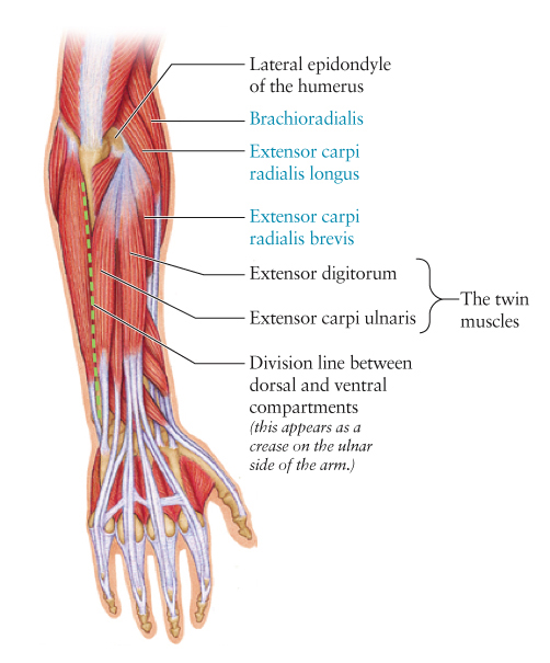
So. When we last left off in our forearm saga, we were looking at, among other structures, the "twin muscles," two muscles that look very much alike and run directly down the dorsal side of the forearm. Their similarity to one another as well as their central location on the dorsal forearm make them among the easiest to identify in this area.

The last three muscles we'll cover on the dorsal forearm can be found just radial to the twins-- meaning closer to the thumb side of the arm compared to the centrally located twins. It's no coincidence that each of these three muscles have the root "radial" in their names. As well as indicating that these muscles are found on the radial side of the arm, this root also tells us that these muscles pull the hand toward that side. This movement is known as abduction of the hand. Hence the fact that radial side arm muscles tend to abduct.


The three muscles we'll be looking for today are labeled in blue on this diagram. Notice that they are closest to the radial (thumb) side of the hand. And notice that all their names have "radial" somewhere in the name. 

Of these muscles, the most radial is brachioradialis. This muscle starts way up on the upper arm (or brachium, hence the root brachio in its name) and travels distally, along the radius, towards its insertion on the styloid process of the radius (a bump at its distal end.) This muscle, despite its certified membership in the dorsal forearm compartment, can actually be seen more clearly from the ventral side. So we won't see much of it today in our photographs.

Running right between brachioradialis and  extensor digitorum (one of the twin muscles) we see two muscles with very similar names: extensor carpi radialis longus and extensor carpi radialis brevis. Can you guess, by looking at these names, what these muscles have in common and what they don't?

Let's look at their names: Both names contain extensor carpi radialis, which means extensor of the wrist (carpi) on the radial side of the arm (radialis.) So these are attributes of both muscles. But how do we distinguish them from one another? The qualifiers tacked on to the end of each name tell us! Yes! One of these muscles is longer than the other. Extensor carpi radialis longus is the longer of the two, and extensor carpi radialis brevis is the shorter. (Brevis is Latin for short, or brief.) Knowing this, you should be able to tell one of these muscles from the other, as one is clearly longer than the other. In addition, it's helpful to know that extensor carpi radialis brevis lies right next to extensor digitorum and it tends to sink in rather than stand out when the dorsal forearm muscles show on the surface of the body.

Extensor carpi radialis longus is sometimes identified by its unique shape. It originates just proximal to the lateral epicondyle of the humerus, higher up on the arm than the origin points of the twin muscles. Also, unlike the twin muscles, extensor carpi radialis longus take a sharp turn where its muscle body meets its long insertion tendon. So its muscle body (which is its most visible part on the surface of the body) appears at an oblique angle on the upper dorsal-radial forearm. This is the only dorsal forearm muscle that lies at such an angle.


Key: Tlat: triceps, lateral head; Tten: triceps tendon; BrR: brachioradialis; ECRL: extensor carpi radialis longus; ECRB: extensor carpi radialis brevis; LE: lateral epicondyle; Anc: anconeus; OP: olecranon process; ED: extensor digitorum; EDM: extensor digiti minimi; ECU: extensor carpi ulnaris; FCU: flexor carpi ulnaris; APL: abductor pollucis longus; EPB: extensor pollucis brevis; EPL: extensor pollucis longus

The photo above shows a dorsal forearm and an abducted hand. Because radial side muscles abduct the hand, they will stand out more in this position than in any other. The easiest one to spot is extensor carpi radialis longus, as it stands out clearly right next to the lateral epicondyle. Notice its oblique course compared to the surrounding dorsal forearm muscles. Notice also that it and brachioradialis originate on the upper arm (unlike the twin muscles that originate at the lateral epicondyle of the humerus.)

At the distal end of the arm, you may also notice three smaller muscles, abductor pollucis longus, extensor pollucis brevis, and extensor pollucis longus. While these muscles stand out clearly here, they often don't. We will look at them more closely later. You may remember, however, that we have already observed their tendons (which can be seen at the base of the thumb) in the post on the dorsal hand.

I've also pointed out the lateral head of the triceps and the triceps tendon, structures on the posterior upper arm. We will cover these later, but this photo shows them very clearly.

Now let's take a look at this arm without the structure overlay:


Notice the most obvious radial side muscle is extensor carpi radialis longus. It bulges out more than its counterpart, extensor carpi radialis brevis. Brachioradialis does show fairly well on the surface, but as pointed out earlier, it actually shows more clearly on the ventral side of the forearm. Brachioradialis is one of the dorsal forearm muscles that can't decide which side it wants to be on; although it's technically in the dorsal compartment, it tends to peek around to the ventral compartment, and although it technically belongs to the extensor muscle group, it does facilitate some flexion of the wrist as well. In any case, we don't see it very clearly on the dorsal side of the arm, nor to we see extensor carpi radialis brevis very clearly (other than in an exceptionally defined individual.) As such, if the hand is abducted, the muscle we want to look for (and to be sure to draw!) is extensor carpi radialis longus.

So let's look for this muscle in a few more images...


In the photo above, we can see three structures very clearly. (Well, four, if you count the extensor digitorum tendons on the back of the hand.) We can see extensor digitorum (the more radial of the twin muscles), the lateral epicondyle of the humerus, and the extensor carpi radialis longus muscle. Notice again how ECRL runs more obliquely than its neighboring muscles and how it originates higher up on the arm than the twin muscles.


The extensor carpi radialis longus muscle is softer in this photo but still visible because the hand is abducted. We can also see the lateral epicondyle, anconeus, and both extensor digitorum and extensor carpi ulnaris. The degree to which the dorsal forearm structures are visible on the surface of the depends on several variables. These include but aren't necessarily limited to: the tone of the muscles, the amount adipose tissue overlying the muscles, the position of the arm and the degree of muscle contraction, the age of the individual (as it relates to skin thickness) and even the light source and the amount of contrast in the values of the structure.

Well, we've covered just about everything we can on the dorsal forearm, with the exception of the radial thumb muscles. Perhaps that can be our epilogue? But I'm going to put the forearm aside for awhile and move on to something different. I'm thinking maybe the posterior torso muscles or something with the thigh. We'll see. As always, suggestions are welcome!

Thanks to my forearm models, Christian, Jessica, and Jeff. I couldn't write this blog without your willingness to stand around and do funny poses for me.

Thursday, July 28, 2011

The Dorsal Forearm, Part 2: Which Side Are You On, Anyway?

Now that we’ve established a method for identifying the two compartments of the human forearm, let’s look at the dorsal compartment a little more closely. The dorsal side of the forearm is the "top" side-- the side that usually faces upward and is generally darker and hairier (due to more melanin and a greater number of hair follicles.) The muscles on the dorsal side of the arm (unlike those on the ventral side) are arranged in a single layer, so they all lie directly under the surface of the skin. This means most of them can be seen pretty clearly on a well defined individual, although their relative visibility will depend on the position of the hand. The muscles in this compartment are all part of the extensor/supinator muscle group, which means they either extend the wrist or fingers or supinate the forearm, or both.

When considering which muscles will show in a given position, we want to keep in mind what the muscles in a specific area tend to do. We are looking at the extensor/supinator group today, so it's safe to assume that these muscles can be seen more clearly when we are supinating the forearm (which means turning the palm upward) or extending the wrist or fingers (which means opening them up.) It's also helpful to remember that muscles on the radial (thumb) side of the arm tend to pull the hand in the direction of the radius (or abduct it) and muscles on the ulnar (pinky) side of the hand tend to pull the hand in the direction of the ulna (or adduct it.) As such, radial side muscles tend to show more when the hand is abducted and ulnar side muscles tend to show more when the hand is adducted. Knowing these four movements will help us to remember which hand positions will make which muscles stand out.

So... here is an overview of what we'll cover today:


When identifying muscle shapes on the forearm (or anywhere else on the body, for that matter) it’s best to locate the most obvious structures first and then find the other structures based on their relationships to the former. Today we'll look at the ulnar (pinky) side muscles, because I think they're easier to find first. Once we've established their location, we'll go on to the radial (thumb) side in the next post.

When becoming oriented on the dorsal forearm, I usually begin with what I like to call the twin muscles-- two muscles the seem to look more alike than any of the others. They are often the first to make themselves evident when looking at the dorsal forearm. The twin muscles are extensor digitorum and extensor carpi ulnaris. They both originate at the lateral epicondyle of the humerus (the bony bump on the lateral side of the elbow) they are about the same width, and they both run straight down the dorsal side of the forearm, toward the hand. Find them below in both the anatomical rendering and the photograph, and notice how similar they are.

The dorsal forearm "twin" muscles, extensor digitorum and extensor carpi ulnaris, look very much alike. We can distinguish one from the other by remembering that extensor carpi ulnaris is closer to the ulnar (pinky) side of the arm. 

Please note that the elbow position in the photo does not exactly match that in the diagram. But for our purposes, we can see what we need to see: The twin muscles and the furrow where the two muscle groups meet (shown with a dashed line.)

OK, so now that we’ve found the twin muscles, the next step is to distinguish them from one another. This is easy. The twin muscles are extensor digitorum and extensor carpi ulnaris. It makes sense that, of the two, extensor carpi ulnaris is closer to the ulnar side of arm. It actually lies right next to the ulna (whose location is designated by the furrow on the forearm, marked with a dashed line in the photo.) Once we've positively identified extensor carpi ulnaris, we can assume the other twin must be extensor digitorum.

The next muscle we'll locate is extensor digiti minimi. As its name tells us, this muscle extends the small finger (digiti minimi is Latin for smallest finger.) Extensor digiti minimi is very easy to locate because it's very narrow and lies right between the twin muscles. On the anatomical diagram, you can see that the tendon of this muscle is headed straight for the smallest digit, where it will insert on its dorsal side. This tendon is often visible on the dorsal hand. You can see an example of this in an earlier post called The Dorsal Hand: The Dorsal Foot's Better Looking Sibling. The image below demonstrates the location of both extensor digiti mimimi and the last muscle we'll cover today, anconeus.

Location of extensor digiti minimi and anconeus. 

Anconeus is somewhat easy to remember, because it's the shortest muscle in the extensor/supinator group and has the shortest name. It is triangular in shape and can be found running from the lateral epicondyle of the humerus to the proximal end of the ulna. As mentioned in the last post, it can also be found by following the crease along the ulnar side of the arm (the one that divides the two forearm muscle groups) proximally (which means upward, toward the body) until it suddenly ends. When it ends, you'll see the triangular shape of anconeus.

Here is one last image to help you visualize more clearly the four muscles we covered today. This is a repeat from part 1 and shows outlines of all the muscles in the dorsal compartment. Look closely at those we covered today: extensor carpi ulnaris, extensor digitorum, extensor digiti mimini, and anconeus. Ignore the rest for now; we'll get to them soon.


Next time we'll find the last three muscles in the dorsal forearm, those that run along the radial side. They are a little more difficult to spot, but if were start by locating the twin muscles, everything else falls into place pretty clearly.

There are also three muscles at the distal end of the dorsal forearm, but those move the thumb around, and most of what we see of them is their tendons, which show up on the wrist. So we'll cover those in a wrist post. Thanks again to my model, Shannen. See you next time!


Tuesday, July 19, 2011

The Dorsal Forearm, Part 1: Compartment Search

While I'm very happy to get to the first dorsal forearm post, let me first apologize for the long hiatus I've inadvertently taken. In addition to teaching at the American Academy of Art, I am also on the part time faculty at the Biomedical Visualization program at the University of Illinois at Chicago, a fabulous graduate program for future medical illustrators, animators, and anaplastologists. Although it's kept me from my blog for the past few weeks, I've very much enjoyed reading and editing many compelling research papers that this year's graduating BVIS students are finishing up. I can't give away their content, but let's just say it's amazing to see what can be done with MRI data and 3D modeling and animation applications!

The dorsal forearm is one of my favorite lectures, as its intricate and complex musculature is unmatched anywhere else in the body. At the Academy, we devote three entire classes to just the forearm; it deserves a slow and thorough explanation. I can't fit even the dorsal side into one post here, so I'll be dividing it up into a few.

Muscles in the limbs are categorized into groups (which are defined by function) that reside in muscle compartments (which are defined by location.) The forearm has two muscle compartments, the dorsal compartment (in which the extensor/supinator muscle group resides) and the ventral compartment (in which the flexor/pronator muscle group resides.) I wish I could say the dorsal and ventral compartments were placed perfectly dorsally and ventrally on the arm, but alas, they are not. Each spills over onto the other side a bit. This means while most extensor/supinators are seen on the dorsal side of the arm, a few can be seen peeking over onto the ventral side, and while most flexor/pronators are seen on the vental side of the arm, a few can be seen peeking over onto the dorsal side. 

Have I lost you yet? Agh, I know this verbal explanation is really dry, so lets try a diagram. Click for an enlarged view if necessary!


Above we see dorsal (top side) and ventral (underside) views of the right forearm. We also see the location of the extensor/supinator muscle group (shown in blue) and the flexor/pronator muscle group (shown in green.) Notice how we see mostly extensor/supinators on the dorsal side, but a few flexor/pronators creep over there. Similarly, we see mostly flexor/pronators on the ventral side, but a few extensor/supinators creep over there. It's like they each know where their home is, but they can't resist peeking around to the other side.

Since the two forearm muscle groups are not clearly divided between the ventral and dorsal sides, we use other landmarks to find their borders. On the dorsal side of the arm, the crest of the ulna (the distal end of the bone that can be seen and felt on the surface of the arm) is the border between the two muscle groups. On the ventral side, the biceps brachii tendon (coming from the biceps brachii muscle on the anterior upper arm) is the border between the two muscle groups.

When observing the muscles of the forearm, the best way to become oriented is to first figure out where the two muscle groups (and the borders between them) are. As such, when observing the dorsal forearm, the first structure we want to identify is the ulnar crest. This actually doesn't look like much on the surface, but it's easy to find A) because we can usually feel it, and B) because it merges with a nice little furrow at its proximal end.


In the above photograph, the arrow is pointing to the crease along the ulnar crest. Everything above this in the photograph is the extensor/supinator group, and everything below this is the flexor/pronator group.


In this photo, the dashed line follows most of the furrow along the ulna distally until we reach the head of the ulna (the little bump on the pinky side of the wrist. The furrow stops suddenly at its proximal end because at that point we run into the anconeus muscle, a small triangular muscle between the lateral epicondyle of the humerus and the olecranon process of the ulna (what we normally think of as the elbow.)


The area in blue (above the red dashed line) is the extensor/supinator group. The area in green (below the red dashed line) is the flexor/pronator group. Muscles are outlined and labeled:  BRR (brachioradialis); ECRL (extensor carpi radialis longus); ED (extensor digitorum); EDM (extensor digiti minimi); ECU (extensor carpi ulnaris); Anc (anconeus); and FCU (flexor carpi ulnaris) which is the only muscle from the flexor pronator group that can be seen from this view.

In the above image, we can still see the division between the two muscle groups (shown with a red dashed line) but we can also see outlines of muscles within each compartment. Notice how the division line runs right along the ulnar crest and ends distally at the head of the ulna. Notice also how the proximal end of the division line ends at the triangular anconeus muscle.

Now that things are a little more clarified, lets revisit the unembellished arm photo and take another look at the muscles shapes.


So I think that's all we'll cover for today. Keep in mind that these early posts tend to show the body in odd positions because we are still covering straightforward anatomy, and these positions are meant to help pronounce all structures in a given area. Later on we'll observe the arm (and the rest of the body) in more natural poses and note what structures are seen then. 

Dorsal forearm part 2 to come soon! At that point we'll look more closely at the individual muscles and learn to tell them apart. Thanks to my forearm model, Shannen!

Tuesday, May 3, 2011

The Ventral Forearm: What are those Tendons?


While the ventral side of the forearm is not exactly less complicated than the dorsal side, it appears less complicated on the surface because so few of its structures show clearly on the surface of the body. Compared to the dorsal side of the forearm, the ventral surface is smooth and uncomplicated. The ventral side of a vertebrate is generally considered its "underbelly"-- paler and less hairy than its dorsal counterpart because of fewer hair follicles and less melanin production.

But the distal end of the ventral forearm (the end closest to the wrist) does have a few prominent surface landmarks. I write "a few" instead of a specific number because the number depends on the individual; one of the tendons that's often seen at this location comes from a muscle that is actually missing in 12-15% of the human population! And, if that muscle and tendon are missing, some deeper tendons may or may not show!

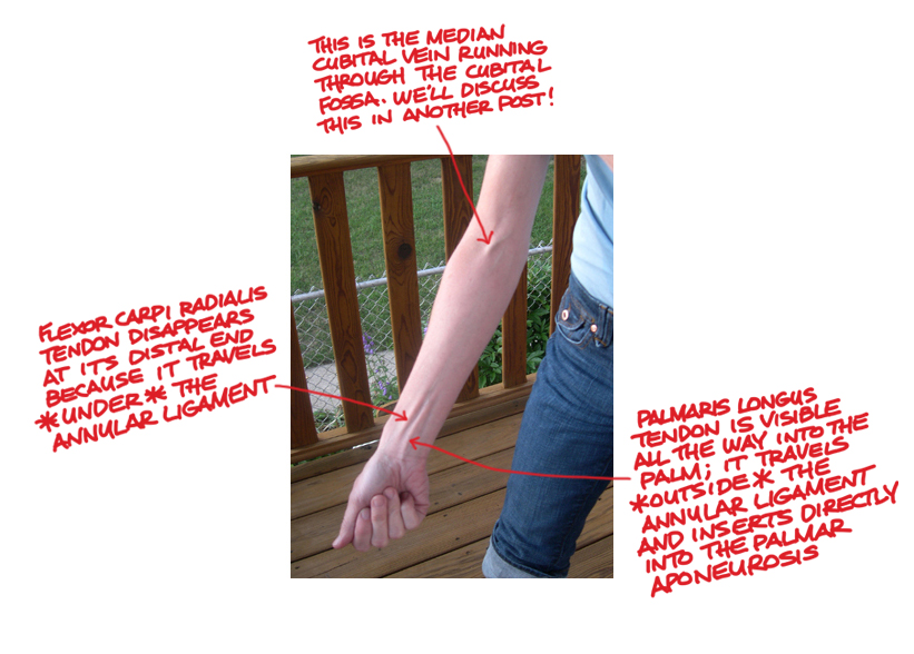
Let's back up a little. In most cases, there are two fairly visible tendons down the approximate center of the ventral wrist. (Their degree of visibility also depends on genetics, hand position, and temporary body variations such as those seen in water retention.) Those two tendons come from the palmaris longus muscle and the flexor carpi radialis muscle. They are shown in the illustration below. We can tell this is a ventral view of the forearm because we can see the palmar aponeurosis (a thin, tendinous sheath that is only on the palmar side of the hand) and because, um... there are no fingernails!


If both the flexor carpi radialis tendon and the palmaris long tendon are visible, it's easy to tell one from the other; the flexor carpi radialis tendon is more toward the radial (thumb) side of the arm, as its name implies. Once that's been established, one can deduce that the other tendon is most likely that of palmaris longus.

The palmaris longus tendon is also more superficial than the flexor carpi radialis tendon because it runs outside the annular ligament, while all the other wrist tendons run deep to it. The annular ligament is a ring-like ligament ("annular" is Latin for "ring-like") that wraps around the wrist like a bracelet and retains the position of the tendons that run from the forearm and into the hand. After surpassing the annular ligament, the palmaris longus tendon inserts directly into the palmar aponeurosis and tenses it to help strengthen the grip.

In some cases, one or both of these tendons are difficult to see, especially when the hand is relaxed. Because both of these muscles are flexors, you can force them to stand out more by flexing the wrist and tensing the hand into sort of a claw shape.

In the photo below, you can easily see both ventral forearm tendons and tell them apart. First of all, flexor carpi radialis is the more radial of the two (meaning it is closer to the thumb side.) Second, palmaris longus can be seen more clearly where the wrist meets the hand, because at that point, flexor carpi radialis is traveling under the annular ligament and palmaris longus is not.


But because palmaris longus is missing in 12 to 15% of the human population, we must also consider what the wrist looks like if there is no palmaris longus muscle. If the palmaris longus muscle is missing, you can usually still see the tendon of flexor carpi radialis. Ulnar to that, where the palmaris longus tendon would normally be seen, you might see nothing but smooth skin. Or you may see some less pronounced tendons, which would most likely be those of flexor digitorum superficialis, which lies deep to everything we've talked about so far. Flexor digitorum superficialis is Latin for "superficial flexor of the fingers," which implies there is also a deep flexor of the fingers (flexor digitorum profundus.) But that muscle is very deep and there is usually no evidence of it on the surface.

In my Anatomy class at the American Academy of Art in Chicago, we always end week 11 ("forearm week") with all my students making claw hands and checking to see whether they have the palmaris longus muscle. As expected, most students have it, a few don't, and occasionally one or two have palmaris longus in one forearm but not the other. But there is one occurrance of which I've found I can be almost 100% sure: Those students who do not have the palmaris longus tendon always seem bothered by this fact, and some actually refer to themselves as freaks! Let's make this clear once and for all: You are not a freak if you don't have the palmaris longus muscle! Distinctive, yes, but not a freak.

Do you have the palmaris longus muscle? See if you can tell and let me know. I welcome your photos, comments, and questions!

Until next time,
Kristin