Showing posts with label forearm muscles. Show all posts

Thursday, June 6, 2013

A Beautiful Exaggeration: More Student Forearm Paintings

I promised I'd post more student work this month, so here we go. Last time we saw Izzy Carranza's lovely spine sculpture, so this time I thought we'd check out some beautiful watercolor paintings by my student Jeff Sant, also in the Spring 2013 Advanced Anatomy class.

You know I love arm anatomy, right? The disproportionate amount of arm posts on this blog sort of gives it away. So it was a lot of fun working with Jeff on this project, in which he drew a somewhat exaggerated arm outline and, in two separate paintings, placed bony and muscular anatomical structures in it. Let's first look at the muscle painting. Please do yourself a favor click on this lovely painting for a full size view.

Watercolor painting of hand, dorsal forearm, lateral arm, and
posterior shoulder musculature by Jeff Sant.

One of the cool things about this painting is its demonstration that even exaggerated anatomy can and should still take its cues from proportional anatomy. Yes, the hand might be larger than usual, yes some of the muscle shapes are unusually pronounced. But that's cool. We still want it to be based on what we've learned from more realistic anatomy examples. Exaggeration doesn't work unless it's based on reality. It's all about comparison.

Another thing I like about this image is the lovely colors and textures of different body tissues. The bones appear solid, calcified and a bit rough, the tendons appear fibrous and flexible, and the muscles appears meaty and striated. Too often paintings of anatomy look like paintings of plastic anatomy models. These tissues look alive.

So are you wondering what you're looking at in this painting? I thought you might be, so let's label it. Again, please click to enlarge.

Muscles are labeled in black. Bony landmarks are labeled in blue.

I haven't labeled everything in this image, but I tried to address anything you would or could see on the surface of the body. As usual surface appearance of structures depend on many variables, including body position, the amount of adipose tissue, and lighting in the room. But everything labeled here could be seen under the right circumstances. Muscles are labeled in black and bony landmarks are labeled in blue. Notice how many of the forearm extensors (including the extensor carpi ulnaris muscle, the extensor digiti minimi muscle, the extensor digitorum muscle, the extensor carpi radialis brevis muscle, and the anconeus muscle) all original on the lateral epicondyle of the humerus.

You can read more about these muscles in the following posts:

The Dorsal Forearm, Part 1: Compartment Search

The Dorsal Forearm, Part 2: Which Side Are You On, Anyway?

The Dorsal Forearm, Part 3: The Final Chapter

The Dorsal Forearm: One Last Encore

The Deltoid Area: Soft Shoulder and Varied Terrain

The Posterior Torso Muscles: Let's Go Back In Time

The Posterio Torso Muscles, Part 2: Under the Radar

Quick Forearm Study: My Pal Rich


Yeah, I told you I liked the forearm.

Soooo, let's go on to the bones of the arm. Yes, Jeff also did a lovely painting of just the bone anatomy. Here it is.

Watercolor painting of the skeletal structure of the hand, dorsal forearm, lateral arm,
and posterior shoulder by Jeff Sant.


Another very cool thing about these two paintings is that they line up accurately with one another. The same outline was used for each, and the bones of this one are arranged so they align perfectly with the muscles and bony landmarks of the other. In addition, while this painting allows us to see the complete structure of the bones, it also allows us to see why certain features of bones are more visible on the surface. For example, we can see the lateral epicondyle of the humerus because, although many forearm extensors originate there, none of them obscure it. And that epicondyle is just under the surface of the skin. The rest of the humerus, however (other than the medial epicondyle, which can't be seen from this view) is completely obscured by muscle tissue.

Now let's look at a labeled version of this painting to see which bone features are surface landmarks:

All bones are labeled. Those features that appear as surface landmarks are labeled in green.

I've labeled all the bones, but the features that are not obscured my muscle and as such appear as surface landmarks are labeled in green.

You can read more about the elbow joint in The Elbow Joint, Part 1: Anterior View, Supine Position.

One last thing I'd like to say is that my favorite part of teaching this class (other than all the lovely work that comes in at the end) is the process of working with students to figure out the great anatomy puzzles that we're presented with when they choose their final assignments. All of these pieces are so complex that they take many rounds of roughs and revisions before finalization. When getting together the work for this post, I found a nice little image that demonstrates part of this process-- a sketch from the beginning phases of Jeff's muscle painting. Often sketches are sent to me via email throughout the school week so I mark them up in Photoshop to try to help clarify things. This process, both in class and out of class, is one of my favorite parts of teaching. Here's the sketch with some color coded adjustments:


Well, I think that's enough for this time. Many thanks to Jeff for letting me use his work. Please check out more of Jeff's work here.

Next time I'll be featuring more student work—that of the awesome Justine Herrera. You can see a shot of one of her pieces here. My bad phone shot absolutely doesn't do her work justice, but I will soon get higher quality images of it to share with you. See you then.

Wednesday, May 22, 2013

Up Close and Personal: Let H.A.F.A. Diagram YOUR Anatomy

This is quite possibly one of the stranger questions you've been asked, but have you ever wanted to have a photo of yourself— your neck, your back, your arm, your foot— diagrammed out anatomically like the photo below? If so drop me a line at kristin@gm-studio.com. Include any photos you have in mind and we'll discuss the options. Then I will diagram it to your specifications for $25 and up, depending on the area to be diagrammed and the detail level of the rendering. When the diagram is complete, I'll send you the finished digital file or FTP it to the service bureau of your choice for digital output. More information to come, so keep reading:

One of my first posts on this blog (and one of my favorites) was The Anterior Neck: Theme and Variations, in which the visible structures on the anterior neck and their variability were examined. As much as I wanted to write about the the beautiful and elaborate anatomy in this area, I could not find an appropriate image that showed everything I wanted to show. So I ended up taking a shot of my own neck and diagramming it out.


While it's an amateur photo taken with an inexpensive camera, I was able to choose position, lighting, and the structures that would show most. It serves its purpose. Since then I've typically relied on shooting my own photographs, both for this blog and for my book. Soon after this post was published, a friend saw it and subsequently sent me a photo of his beefy arm, asking if I'd diagram the muscles out for him.

Since then, I've been getting more and more requests to diagram muscles and bony landmarks on personal photos, not only from friends and relatives, but from readers of this blog. This is a lot of fun and it seems to be gathering momentum, so thought I may as well make it official and offer it to everyone.

Sooo... Have you ever wondered exactly which muscles and bones you're seeing on the surface of your own body? Well, let me diagram them for you. Just send me a clear, high resolution (at least 300ppi) image of the area in question (um, no private parts, please) and let me know how detailed you'd like the diagram. (See below for examples.) Prices go from $25 up, depending on the complexity of the area and the detail level requested.

The most detailed example would be the fully rendered anterior neck image shown above. Simpler diagrams would look more like the following, which just simple outlines, color coding, and labels:

This is the diagrammed dorsal forearm of my student Shannen.
Bones, muscles, and compartment divisions are color coded and labeled.

Another example of this level of detail can be seen here:


I can also take your color or grayscale photo and make it into a sepia image before laying in the diagram, as shown below:


The image above shows the sepia option, but not much a diagram. I'm happy to complete any level of rendering over an image that I've converted to sepia.

A final option is to show only a specific few muscles, and/or simply labeling visible muscles, as in the diagram below. This option, in which only a few structures are diagrammed, can also be rendered with greater detail, like that in the anterior neck diagram up above.


The above image is a much simpler forearm diagram. Only three of the extensors are drawn in, along with the dorsal hand tendons of one muscle. The lower image has only muscles labeled, with no diagramming at all. This wouldn't be as much fun for me, but I'd be happy to do it!

I've shown mostly arms here, but legs, feet, hands, abdomen, back, and head are all fine. I welcome a challenge! Just be sure the image you send is clear and high resolution, and that some level of surface landmark detail can be seen.

For a time frame and quote, send images to kristin@gm-studio.com.

And feel free to contact me at this address with any other questions!

Tuesday, May 3, 2011

The Ventral Forearm: What are those Tendons?


While the ventral side of the forearm is not exactly less complicated than the dorsal side, it appears less complicated on the surface because so few of its structures show clearly on the surface of the body. Compared to the dorsal side of the forearm, the ventral surface is smooth and uncomplicated. The ventral side of a vertebrate is generally considered its "underbelly"-- paler and less hairy than its dorsal counterpart because of fewer hair follicles and less melanin production.

But the distal end of the ventral forearm (the end closest to the wrist) does have a few prominent surface landmarks. I write "a few" instead of a specific number because the number depends on the individual; one of the tendons that's often seen at this location comes from a muscle that is actually missing in 12-15% of the human population! And, if that muscle and tendon are missing, some deeper tendons may or may not show!

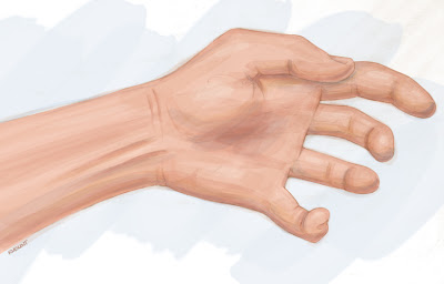
Let's back up a little. In most cases, there are two fairly visible tendons down the approximate center of the ventral wrist. (Their degree of visibility also depends on genetics, hand position, and temporary body variations such as those seen in water retention.) Those two tendons come from the palmaris longus muscle and the flexor carpi radialis muscle. They are shown in the illustration below. We can tell this is a ventral view of the forearm because we can see the palmar aponeurosis (a thin, tendinous sheath that is only on the palmar side of the hand) and because, um... there are no fingernails!


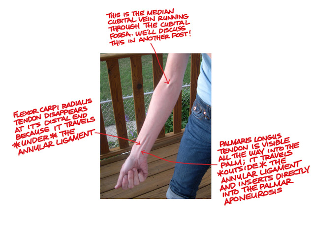
If both the flexor carpi radialis tendon and the palmaris long tendon are visible, it's easy to tell one from the other; the flexor carpi radialis tendon is more toward the radial (thumb) side of the arm, as its name implies. Once that's been established, one can deduce that the other tendon is most likely that of palmaris longus.

The palmaris longus tendon is also more superficial than the flexor carpi radialis tendon because it runs outside the annular ligament, while all the other wrist tendons run deep to it. The annular ligament is a ring-like ligament ("annular" is Latin for "ring-like") that wraps around the wrist like a bracelet and retains the position of the tendons that run from the forearm and into the hand. After surpassing the annular ligament, the palmaris longus tendon inserts directly into the palmar aponeurosis and tenses it to help strengthen the grip.

In some cases, one or both of these tendons are difficult to see, especially when the hand is relaxed. Because both of these muscles are flexors, you can force them to stand out more by flexing the wrist and tensing the hand into sort of a claw shape.

In the photo below, you can easily see both ventral forearm tendons and tell them apart. First of all, flexor carpi radialis is the more radial of the two (meaning it is closer to the thumb side.) Second, palmaris longus can be seen more clearly where the wrist meets the hand, because at that point, flexor carpi radialis is traveling under the annular ligament and palmaris longus is not.


But because palmaris longus is missing in 12 to 15% of the human population, we must also consider what the wrist looks like if there is no palmaris longus muscle. If the palmaris longus muscle is missing, you can usually still see the tendon of flexor carpi radialis. Ulnar to that, where the palmaris longus tendon would normally be seen, you might see nothing but smooth skin. Or you may see some less pronounced tendons, which would most likely be those of flexor digitorum superficialis, which lies deep to everything we've talked about so far. Flexor digitorum superficialis is Latin for "superficial flexor of the fingers," which implies there is also a deep flexor of the fingers (flexor digitorum profundus.) But that muscle is very deep and there is usually no evidence of it on the surface.

In my Anatomy class at the American Academy of Art in Chicago, we always end week 11 ("forearm week") with all my students making claw hands and checking to see whether they have the palmaris longus muscle. As expected, most students have it, a few don't, and occasionally one or two have palmaris longus in one forearm but not the other. But there is one occurrance of which I've found I can be almost 100% sure: Those students who do not have the palmaris longus tendon always seem bothered by this fact, and some actually refer to themselves as freaks! Let's make this clear once and for all: You are not a freak if you don't have the palmaris longus muscle! Distinctive, yes, but not a freak.

Do you have the palmaris longus muscle? See if you can tell and let me know. I welcome your photos, comments, and questions!

Until next time,
Kristin