The lower leg has three muscle compartments-- the anterior, the posterior, and the lateral. In each of these fall muscle groups, each with its own functional purpose: In the anterior compartment we find the foot extensors and dorsiflexors; in the posterior compartment we find the foot plantarflexors, and in the lateral compartment we find the foot everters. A later post will elaborate on these movements.
Today we'll be discussing the anterior compartment of the lower leg, but only its muscles that actually appear on the lower leg. Some muscles in the anterior compartment, while they lie in the lower leg, don't show up on its surface. Their tendons may show, but they don't surface until they've already reached the foot. Those particular muscle tendons are discussed in The Dorsal Foot: How Do I Love Thee? Let Me Count Your Tendons. The tibialis anterior, it turns out, is the only muscle whose body can clearly be seen on the anterior surface of the lower leg. It's on top of everything else, and it stands completely alone.
DO YOU KNOW WHAT THIS MEANS??? It means that after this long, long, boring introduction, we're only going to cover one muscle today-- the tibialis anterior. As you may remember, I was planning to cover the tibialis anterior muscle last time but quickly realized it was impossible without first going over the lower leg bones; although tibialis anterior stands alone muscularly, its relationship with the tibia is the key to its identification.
So... let's start with an overview of the muscles in this area and their relationship to the bones covered last week in The Anterior Leg, Part 1: The Supporting Cast.
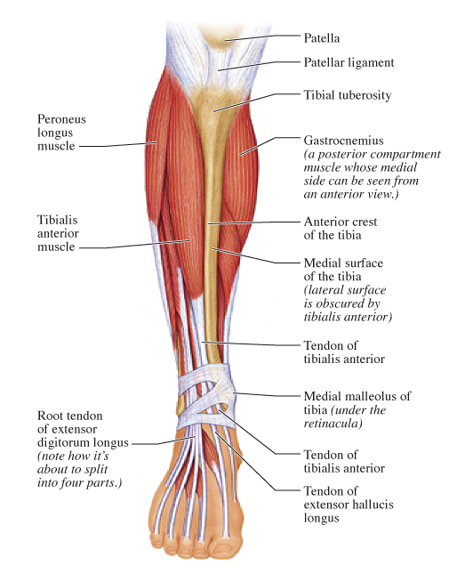 |
| The entire medial surface of the tibis is exposed, but the lateral surface is obscured by the tibialis anterior muscle. |
As we saw in the last post, the tibia is the larger and more medial of the two lower leg bones. There is a long ridge down its anterior side known as the anterior crest. One either side of the anterior crest are two long, flat surfaces. The medial of these (called, um, the medial surface) is completely exposed. It comes right up to the surface of the body, and it's what we colloquially refer to as the shin.
The lateral surface of the tibia is not a surface landmark because it is almost entirely obscured by, YES, the tibialis anterior muscle! This lovely little structure moves the whole foot, and is the only muscle in the anterior compartment to do so. (The other muscles in this compartment move the toes.) Because tibialis anterior is in the dorsiflexor group, it dorsiflexes the foot, or points it upward. This is not typically a very strong or pronounced foot movement, but it is important in making sure our foot is lifted up enough with each step so that we don't drag our toes. Yes, this little muscle keeps us from stubbing our toes. (Well, most of the time, anyway.)
Let's take a look at the tibia and the tibialis anterior's appearance on the lower leg:
 |
| The tibialis anterior muscle and its tendon can be seen very clearly on the surface of the leg when the foot is dorsiflexed and inverted. We can also see the vast surface of the medial tibia, as well as several other bony landmarks. |
Notice how the lateral side of the lower leg appears soft and rounded, while the medial side appears flat and smooth. This is because the lateral side is soft tissue (in the form of the tibialis anterior muscle) and the medial side is the long, wide medial surface of the tibia.
Notice also how the tibialis anterior tendon shows clearly from just below the muscle body all the way down to the medial foot. It's most prominent just over the ankle. Notice also that the tendon of the extensor hallucis longus muscle runs parallel with that of tibialis anterior on the dorsal foot. We can tell one from the other, though, because the tendon of tibialis anterior is wider and more medial, and it surfaces more proximally than the tendon of extensor hallicus longus.
I have also pointed out a few other surface landmarks in the photo above, including certain features of the tibia and some dorsal foot tendons that come from anterior leg muscles whose bodies we cannot see up in the leg.
In case the basic muscular and bony shapes need to be clarified, take a look at this very simple diagram, in which the basic bone and muscle shapes are overlaid onto the photo:
 |
One last thing: Did you notice there is no medial compartment in the lower leg? Although we can invert our foot (turn its sole inward) there is no specific compartment whose function is only this. It makes sense that muscles on the medial side of the leg would invert the foot-- or pull it medially-- but alas, there is no medial compartment. But it turns out a medial compartment is not necessary here, because two other muscles on the lower leg take care of inversion. Gastrocnemius (in the posterior compartment) helps with inversion of the foot, and so does our friend tibialis anterior.
This means tibialis anterior and its tendon really show when we are both dorsiflexing and inverting at the same time (or pointing the foot upward and inward at the same time.) Notice the foot in the photos is held in that position to ensure the best possible view for the camera.
So now we're familiar with our first lower leg muscle compartment. We'll move on to the posterior and lateral compartments in upcoming posts, but I think we might first take a short break from the leg and spend a little time going over the basic terminology of direction and location on the human body. This will help define a great deal of the words used over and over again in these posts. Until then, be sure to thank lonely little tibialis anterior next time you walk without stubbing your toe.
As I said in the wrong post... I've been remiss in blogging etiquette duties, but finally linked to your post. I apologize for any of the crazies it might bring over. :)
ReplyDeleteJENNIFER SENT ME!1!
ReplyDelete~
Anterior thighs and knees are recommended to be done together but in multiple treatments
ReplyDeleteWha! This is great full blog i like this type blog. Deferentially this blog have this quality big cost, special effects, thank for sharing this blog.
ReplyDeleteNonSurgical Facelift
Your article and anatomy images give me the medical terminology to speak to my doctor about my constant pain. Thank you.
ReplyDelete