Writing about the dorsal foot last time got me thinking about the dorsal hand, which despite its completely different outward appearance, is a pretty close relative structurally. For starters, the bones of the hand sit in a curved arrangement so, like the foot, it's convex on its dorsal side and concave on its palmer side. (The palmar side of the hand is also considered ventral, just like the plantar side of the foot.) Because of this arrangement, most of the soft muscle tissue in the hand is either on the palmar side or tucked between the metacarpals. Most of what we can feel on the dorsal side of the hand is bone, and most of what we can see are tendons (along with a few superficial veins.) Like the dorsal foot, the dorsal hand has seven visible tendons (although we don't always see them all at the same time) and only one clearly visible muscle. And finally, the visible tendons in the dorsal hand, like those in the dorsal foot, are named for the structures onto which they insert and for the movements they facilitate.
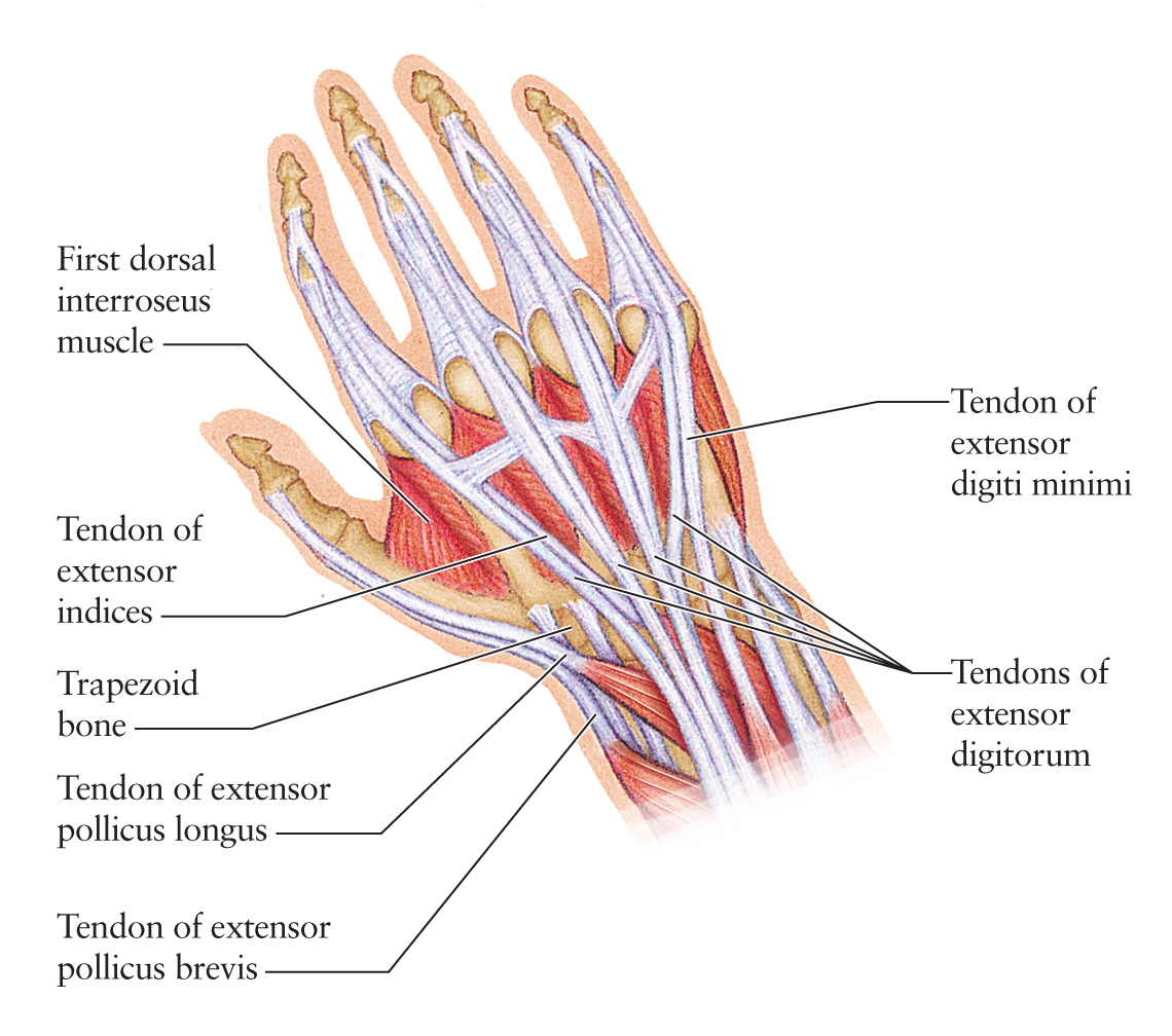
The easiest tendons to identify in the dorsal hand are those of the extensor digitorum muscle. Its name means extensor of the digits, which is why there were also extensor digitorum tendons in the feet. (Toes are digits, too.) But notice the absence of "longus" or "brevis" here; unlike the toes, the fingers have only one extensor muscle, so it needs no further qualifier in its name. The extensor digitorum muscle, which lies in the dorsal forearm, splits off into four individual tendons that insert onto digits II through V (the index finger through the small finger.) The thumb has its own muscles, some of which we'll look at later in this post. It's easy to identify the four extensor digitorum tendons on the dorsal side of the hand because they are clearly heading in the direction of digits II through V. These tendons show most of the time, but more so when the fingers are extended (which means straight as opposed to curled inward) or abducted (which means spread apart.)
The thumb extensors are also pretty easy to spot as well, especially when the thumb is... well... extended. There are two thumb extensors, extensor pollucis longus and extensor pollucis brevis. Pollucis comes from the Greek word pollux. These two tendons are most visible at the base of the hand on the radial (thumb) side. At this point, they rest about one centimeter apart. Of the two, extensor pollucis longus is the more dorsal and, as its name implies, the longer. It inserts all the way on the first distal phalanx. Extensor pollicis brevis is more ventral, and it inserts on the first proximal phalanx. Another tendon, that of abductor pollucis longus, runs right along side the extensor pollucis brevis tendon at this point, but it is difficult to differentiate one from the other. The bodies of all three of these muscles are on the distal end of the forearm, radial side, and you can see them moving around if you extend and abduct your thumb.

Things look a little different on the dorsal hand when we adduct the thumb. Adduction of the thumb is bringing it inward so it tucks up right along the index finger. When we do this, the extensor digitorum tendons are still somewhat visible, but the extensor pollicis longus and brevis tendons disappear almost entirely. And in this position, a whole new structure pops out-- the first dorsal interosseous muscle. Interosseous means "between bones" and these muscles were given that name because they run in between the metacarpals, the bones in the body of the hand. There is a dorsal set of interosseous muscles and a palmar set (which are also between the metacarpals but on the palmar side.) Of all these, the only one that's ever really visible on the surface is the first dorsal interosseous. That means the dorsal interosseous muscle between the first and second metacarpals. When the thumb is adducted, the first dorsal interosseous muscle plumps up like a little pillow just next to the thumb. The more tightly adducted the thumb, the more this muscle shows. At the base of this muscle lies the trapezoid bone, one of the eight small carpal bones at the base of the hand. The trapezoid bone is not always easy to see, but it can be palpated easily.

There are two more elusive tendons on the dorsal hand that can be seen in certain situations. While the extensor digitorum longus muscle extends digits II through V, two fingers on the hand have extensor muscles exclusively their own. The second digit (or digiti indices) has its own private extensor called extensor indices. As you can probably guess, this tendon is most visible when the index finger is extended separately from the others.

The other elusive dorsal hand tendon is that of extensor digiti minimi. This name means "extensor of the small digit" and, you guessed it, it can be seen most clearly when the fifth digit (digiti minimi) is extended separately from the rest. It runs right along side the extensor digitorum tendon heading toward the fifth digit, but the extensor digiti minimi tendon is closer to the ulnar side of the arm.

All this dorsal hand talk has me amped up to write about the dorsal forearm soon. That's the mother of all complex muscular areas, but it's very cool. I've already chosen my forearm models, which was no easy task! I look forward to that and, as always, I'm open to suggestions for other areas to cover.
I'm glad I found this. Thanks so much for your clear description, it's really useful!
ReplyDeleteI'm happy to hear this! Thank you!
ReplyDeletethese pictures and descriptions are very helpful! i'm a med student studying anatomy and this beats a lot of cartoon drawings
ReplyDeleteThank you for this. I agree it is very useful to all aspiring artists :-)
ReplyDeleteThank you. I agree this is very useful to all aspiring artists.
ReplyDelete